What Does Whiplash Look Like on an MRI?
Published: 4/17/2025
MRI (Magnetic Resonance Imaging) is more detailed for soft tissues and may reveal subtle changes that suggest a whiplash injury. Below, we explain what whiplash-related damage may look like on an MRI, using medical terms from radiology reports translated into plain English..
Loss or Reversal of the Normal Cervical Lordosis (Neck Curve)
Under normal conditions, the neck spine has a gentle inward curve called the cervical lordosis (when viewed from the side, the neck bones form a C-shape curvature with the open side toward the back). After a whiplash injury, this curve can straighten out or even reverse into an abnormal slight backward curve. A radiologist’s report might note “loss of cervical lordosis” or “reversal of the cervical lordosis”. In simple terms, the neck that usually curves inward may become unnaturally straight or bowed outward. One medical article notes that after whiplash the cervical spine can straighten or even develop a reverse curvature (kyphosis) due to the trauma. This change is often temporary and is believed to be caused by muscle spasm – the neck muscles tighten up reflexively after the injury, pulling the spine straight as a protective mechanism.
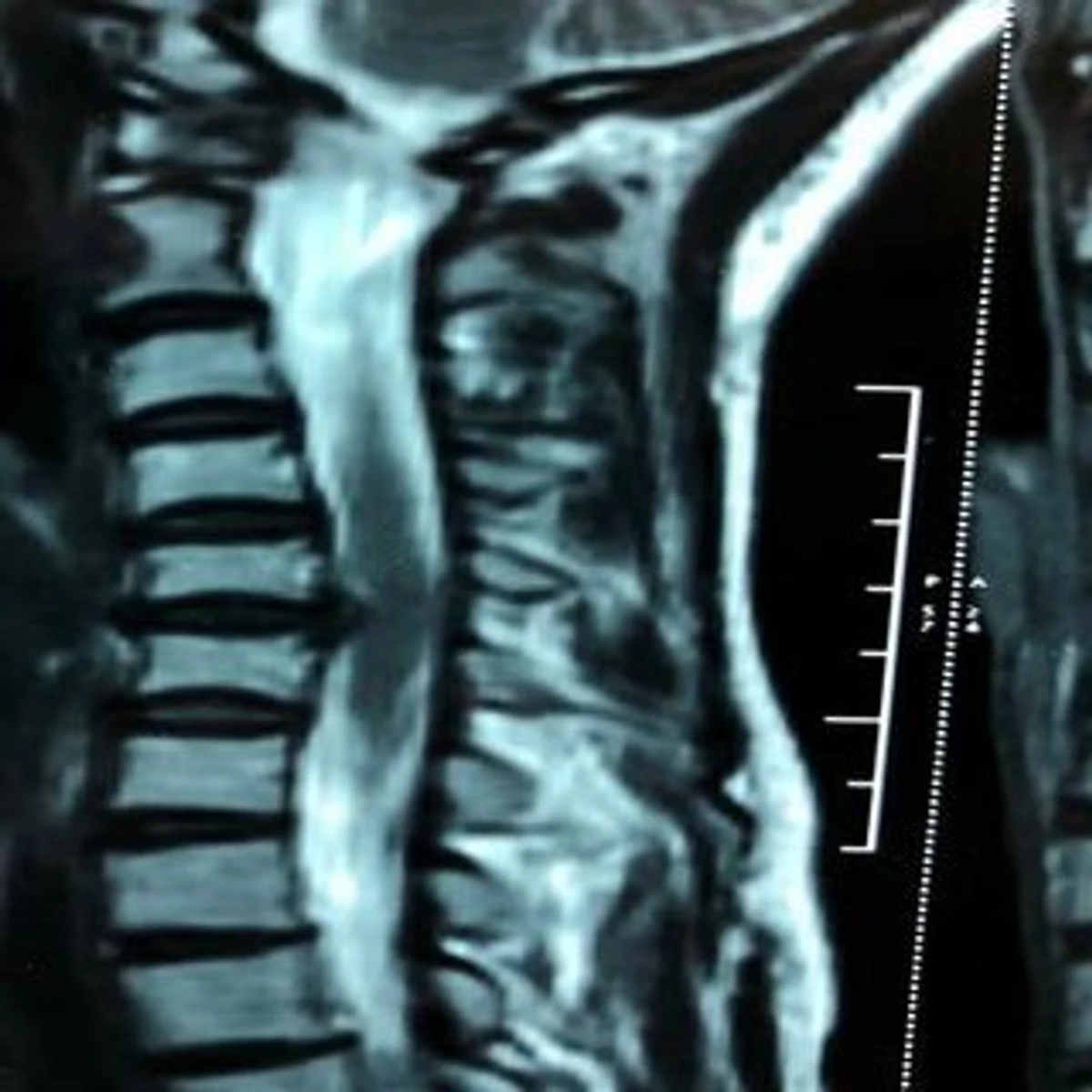
An MRI of the cervical spine (side view) showing a straightened neck curve (loss of lordosis) and a disc bulge at C5-6 pressing toward the spinal cord. The dotted line indicates where a normal forward curve would be. Such an MRI finding illustrates how whiplash trauma can alter the spine’s alignment and damage intervertebral discs.
Medical significance
Losing the normal neck curve is an objective sign that the neck has been injured. While not a specific injury itself, it suggests the neck muscles are in spasm or the spine’s alignment has been disturbed by the whiplash. Patients with a straightened or reversed curve often experience neck pain and stiffness due to this muscle spasm and misalignment. In some cases, studies have found that a sharp reversal of lordosis after trauma can be associated with more severe soft-tissue injury and even predict a higher chance of chronic issues like degenerative changes down the line. Essentially, the body is reacting to the injury by splinting the area (muscles tightening to protect the neck), which shows up on the MRI as an abnormal curvature.
Legal significance
For a personal injury claim, a documented loss of cervical lordosis on an MRI or X-ray can serve as objective evidence that a whiplash injury occurred. It’s a visible, measurable change in the spine’s alignment that wasn’t there before the accident. This can counter any argument that the pain is “all in the victim’s head.” While a straightened neck curve by itself is a relatively subtle finding, it corroborates that the person’s neck experienced an abnormal force. Attorneys sometimes use the loss of lordosis as supporting evidence that the crash imparted enough impact to injure the neck, especially when combined with other findings. It may not be as dramatic as a broken bone, but in legal cases every piece of objective medical evidence helps build the case that the victim sustained a real injury. Documentation of such spinal abnormalities on imaging provides tangible proof of injury, strengthening the patient’s case in insurance claims or legal disputes.
Soft Tissue Swelling and Edema
Whiplash can cause soft tissue swelling in the neck, which might be visible on MRI as areas of edema (fluid). One area doctors pay attention to is the prevertebral soft tissue, which is the tissue in front of the spine. Right after a neck injury, this tissue can become puffy or widened if there’s inflammation or bleeding. In radiology terms, you might see “prevertebral soft tissue swelling” or “prevertebral edema” noted. Even on plain X-rays of whiplash victims, sometimes the only abnormality is an anterior widening of the prevertebral soft-tissue space (meaning the soft tissue band in front of the cervical spine looks thicker than normal). On MRI, this swelling is seen more clearly as a collection of fluid or just a general puffiness in the tissue planes. For example, a medical study of whiplash patients noted MRI findings of fluid and swelling in front of the spine associated with injury to a deep neck muscle.
Medical significance
Soft tissue swelling on MRI is a sign of an acute injury – essentially, it’s evidence of inflammation. If a ligament or muscle in the neck is torn or strained, blood and fluid rush to the area, causing it to swell. On an MRI scan this might show up as a brighter signal in the soft tissue (since fluid has a distinct appearance on certain MRI sequences). For the patient, this swelling itself can cause pain or difficulty swallowing (if severe), but more importantly it flags to doctors that something is injured underneath. Significant prevertebral swelling could raise concern about a hidden structural injury (like a small fracture or ligament tear). Usually in pure whiplash (WAD1-2) the swelling is mild, but its presence reassures the doctor that the patient’s complaints have a physical basis. It also helps in timing – swelling right after an accident shows the injury is acute (new), not an old issue.
Legal significance
In the context of an injury claim, soft tissue swelling on an MRI or other imaging can be a persuasive detail. It’s an objective confirmation of trauma: you can literally see that the tissues are disturbed and inflamed compared to normal. Insurance adjusters often look for objective findings like this. While soft tissue edema alone might not equal a large settlement, in a close case it can corroborate the severity of the impact. For instance, if an MRI report notes prevertebral swelling consistent with a recent whiplash injury, it becomes harder for a defendant to argue that the person just has neck pain unrelated to the crash. It supports the narrative that “something tore or got hurt internally, and here’s the proof – the swelling showing up on MRI.” In short, it adds credibility to the injury claim by backing up the victim’s symptoms with visible evidence.
Disc Bulges or Herniated Discs
The cervical spine is made up of vertebrae (bones) with soft intervertebral discs between them, which act like shock absorbers. A whiplash mechanism can injure these discs. The extreme flexion-extension force may cause a disc to bulge outward or even herniate (rupture), where the inner gel-like material of the disc pushes through the outer layer. On an MRI, a disc bulge or herniation is seen as the disc sticking out beyond its normal boundaries. Radiologists might describe “disc protrusion at C5-6” or “herniated nucleus pulposus” at a certain level of the neck. In lay terms, that means part of the cushion between two neck bones has squeezed out. This can sometimes pinch nearby nerves or indent the spinal cord. MRI is the best tool to detect these disc injuries – in fact, medical guidelines say that patients with whiplash symptoms (especially with arm pain or nerve signs) should undergo MRI to evaluate for disk herniations or other internal damage.
Medical significance:
A herniated or bulging disc in the neck is a significant injury. Discs don’t just bulge for no reason – a whiplash trauma can either create a new herniation or worsen a pre-existing weak disc. If the MRI shows a disc pressing on a nerve root, it explains symptoms like radiating arm pain, numbness, or muscle weakness in the arm or hand. This guides treatment: a small bulge might be managed with physical therapy and pain management, while a large herniation compressing the spinal cord or nerve might require injections or even surgery (such as a discectomy). In whiplash cases, finding a disc injury on MRI elevates the diagnosis from a mild strain to a more concrete structural injury. It’s not uncommon for whiplash patients to have neck pain that persists, and if an MRI reveals a disc bulge/herniation, it provides a clear medical reason. Over time, an injured disc can contribute to chronic pain or accelerated degeneration at that level. Thus, identifying it is important for prognosis – for example, a traumatic herniated disc might lead to early arthritis or need future treatments.
Legal significance:
In personal injury claims, a herniated cervical disc is often a game-changer. This is a well-understood, visible injury that tends to significantly increase case value. It moves the claim beyond “soft tissue” whiplash into the realm of a specific injury that often entails higher medical costs (like surgery or long-term therapy). Objectively, an MRI image of a disc pressing on nerves is hard for insurance companies to refute. It clearly resulted from the accident (especially if the person had no prior neck issues documented). Thus, it helps establish causation: the crash caused a tangible injury. Legally, disc herniations can be classified as permanent injuries in some jurisdictions, which may entitle the victim to greater compensation for pain and suffering and future medical needs. During settlement negotiations or trial, the presence of a disc bulge or herniation on MRI, coupled with a neurosurgeon’s or orthopedic doctor’s testimony, can strongly substantiate the claimant’s complaints of pain and neurological symptoms. It’s important, however, to differentiate acute trauma-related herniation from degenerative (age-related) changes – lawyers may use comparative scans or medical expert opinions to argue the disc damage is acute. The bottom line is that a whiplash MRI showing a disc injury provides solid, objective evidence of a serious injury, often making the personal injury claim much stronger.
Ligament Injuries
Ligaments are tough bands of tissue that connect bones together and stabilize joints. The cervical spine has numerous small but critical ligaments holding the vertebrae in alignment (for example, the anterior longitudinal ligament along the front of the spine, the interspinous ligaments between the spinous processes, and specialized ligaments at the skull-neck junction like the alar and transverse ligaments). A whiplash can stretch or tear neck ligaments, similar to spraining an ankle. On an MRI, ligament injuries can be harder to see than a disc herniation, but they may appear as areas of abnormal signal (bright fluid if torn) or as misalignment of the vertebrae indicating that a ligament isn’t intact. A radiologist might report “ligamentous injury” or “ligament sprain”. Sometimes they infer ligament damage if the spine shows excess movement on flexion/extension X-rays or if there’s swelling where the ligament should be. In more severe cases (WAD grade IV, which involves fracture/dislocation), MRI can clearly show ruptured ligaments (for instance, a torn posterior ligament complex in a serious neck injury). Even in moderate whiplash, doctors will use MRI to look for ligament abnormalities in the cervical region because a partial tear could mean instability.
Medical significance:
Ligament injuries in the neck can contribute to ongoing pain and instability. A sprained ligament (stretched but not fully torn) may not require surgery, but it causes pain, inflammation, and a prolonged healing period. A completely torn ligament in the cervical spine is more serious; if certain key ligaments rupture, the spine can become dangerously unstable (which might necessitate surgical fusion to prevent spinal cord injury). For example, a torn transverse ligament (which holds the first vertebra to the skull) can allow excessive movement between the skull and spine. Thankfully that is rare without severe trauma. More commonly in whiplash, we think of microscopic tears or stretched fibers in the ligaments around the middle or lower cervical vertebrae. These won’t show up dramatically on MRI, but if there’s notable soft-tissue swelling or subtle malalignment, it clues the doctor that a ligament was sprained. Medically, knowing that ligaments are injured reinforces the need for immobilization or careful therapy – you wouldn’t want a patient to aggressively move the neck if it’s unstable. It also explains why a patient might have pain with certain movements long after the accident (ligaments have relatively poor blood supply and can be slow to fully heal). In summary, ligament injuries are an invisible culprit that can make a whiplash more severe than it outwardly appears, and MRI is one of the few ways to get evidence of them (along with special flexion-extension X-ray tests for instability).
Legal significance:
Proving a ligament injury can be a bit tricky in a legal case because it may not be as obvious on imaging, but it’s still very important. If an MRI does show a clear ligament tear or instability, that is strong evidence of a significant injury. Even if it doesn’t appear clearly, a doctor’s testimony that certain MRI signs suggest a ligament sprain can support the claim. From a claims perspective, ligament damage moves the injury beyond a simple muscle strain; it indicates the accident caused structural damage to the neck’s support system. Insurance companies are aware that ligament injuries can lead to chronic neck problems (for example, persistent neck pain or future risk of arthritis due to the loosened support). Thus, when documented, it can increase the settlement value. Also, if the patient had to wear a cervical collar or needed future fusion surgery because of an unstable ligament, those are tangible damages attributable to the wreck. In front of a jury, explaining a “torn ligament in the neck” resonates because people associate torn ligaments with serious injury (just as a torn ACL in the knee is considered serious). One challenge is that defense may argue many ligament findings are subjective or age-related, so having imaging evidence (even subtle, like swelling at the ligament site) and a clear connection to the accident (acute onset of symptoms) is key. Overall, recognizing ligament injuries in whiplash cases is medically and legally significant – it underlines that the victim didn’t just suffer temporary pain, but sustained a real anatomical injury that might have lasting consequences.
Muscle Strain and Spasm Injuries
Whiplash nearly always involves some degree of muscle strain in the neck. During the rapid whipping motion, the neck muscles (such as the sternocleidomastoid, trapezius, and deep neck flexors/extensors) can be overstretched or even sustain small tears. This is essentially a muscle strain (or a tendon strain) similar to what happens if you pull a muscle, but caused by trauma. It also goes hand-in-hand with muscle spasm, which is the muscles tightening up after the injury (as discussed with the loss of lordosis). On MRI, purely muscular injuries can be subtle, but they may appear as areas of edema within the muscle tissue. Radiologists might note “muscle edema” or “muscle strain” if they see it. In some cases, especially if the MRI is done soon after the injury, there could be small tears or bleeding in the muscle visible. One study noted that MRI of whiplash patients, when done within days, can show damage to neck muscles (and even shoulder muscles) as areas of swelling or internal bleeding. In practice, MRI is not always done immediately for whiplash, but if it were, you might see things like a strained muscle with fluid around it or a torn muscle fiber. More commonly, the evidence of muscle injury on MRI is indirect (the straightening of the curve, as muscles in spasm pull the spine straight, or general soft tissue swelling).
Medical significance:
Muscle strains by themselves will usually heal with time, physical therapy, and rest. However, they are responsible for a lot of the acute pain and stiffness after whiplash. When you hear terms like “neck sprain/strain”, it refers to ligament (sprain) and muscle/tendon (strain) injuries. If an MRI specifically mentions a muscle injury (for instance, “edema in the paraspinal muscles at C5”), it confirms the source of pain. Sometimes patients worry that something is “out of place” in their neck, but often the pain is from bruised and overworked muscles. Knowing which muscles are affected can tailor therapy (targeted exercises or trigger point injections). Additionally, if muscle edema is seen on MRI, it indicates the scan was done fairly acutely and it reinforces that the whiplash was severe enough to cause muscle damage (not just a minor tweak). Over the longer term, severe whiplash can lead to muscle deconditioning – interestingly, chronic whiplash patients sometimes show fatty deposits in certain neck muscles on MRI, meaning the muscle has atrophied or healed with scar tissue. This can happen if the injury was bad and the muscle wasn’t used normally for a long time. So muscle findings on MRI can also be a clue in chronic cases (though our focus here is acute whiplash changes).
Legal significance:
Muscle strain might be considered the more “minor” end of whiplash injuries, but it’s still important in personal injury cases. Early on, insurance companies often dismiss whiplash as just muscle soreness with no proof. If an MRI or medical exam notes a specific muscle injury, it pushes back against that narrative. It shows that “yes, the muscles were actually damaged.” However, because soft tissue injuries like muscle strains don’t always show up clearly on imaging, the absence of a noted muscle injury on MRI doesn’t mean the person wasn’t hurt – it just might be beyond MRI’s resolution or the scan was done after the acute phase. When muscle injury does appear on MRI, it is helpful corroboration. For example, if an MRI report confirms swollen neck muscles consistent with strain, a claimant’s report of neck pain is objectively validated. Legally, muscle injuries alone might not yield large compensation (since they usually heal), but they contribute to the overall picture of the injury’s extent. A pattern of muscle spasm noted in medical records over time, despite therapy, can also justify why the person had discomfort for weeks or months. In sum, while a muscle strain may be considered a “soft tissue” injury, having it documented by MRI (or even by a doctor’s physical exam findings of spasm) still plays a role in demonstrating the legitimacy and immediate impact of the whiplash.
Why MRI Findings Matter for Whiplash Victims
The findings on an MRI give doctors a window into the actual physical damage caused by whiplash. This guides treatment and helps validate the patient’s symptoms. For example, if your MRI shows a herniated disc pressing on a nerve, your doctor knows that more aggressive treatment might be needed (like referring to a spine specialist or considering injections or surgery) compared to a case with only mild muscle strain. MRI findings can also alert the doctor to any potentially dangerous injuries (like an unstable spine from torn ligaments, or a spinal cord compression that could worsen without care). Even when MRI results are “normal” or only mildly abnormal, that information is useful to rule out serious pathology. It’s reassuring to both patient and doctor if no major structural damage is seen – then the focus can be on conservative care for the soft tissues. On the other hand, when objective damage is seen, it validates the need for continued medical care. In some cases, it might explain why a patient’s recovery is taking longer. Overall, MRI results combined with the patient’s symptoms and exam findings allow for a targeted rehabilitation plan. They also serve as a baseline: later on, if there are degenerative changes in the spine, doctors can compare to the post-accident MRI to see if the whiplash contributed to any progression of damage.
Legal/Insurance Perspective:
In personal injury and insurance claims, objective evidence is king. MRI findings provide exactly that – concrete evidence of injury. Whiplash claims sometimes get a bad reputation because the injuries are invisible; insurance companies may unjustly label them as exaggerated. But if your MRI shows tangible issues (loss of the normal curve, a disc injury, etc.), it becomes much harder for an insurer or opposing attorney to argue that you weren’t really hurt or that it’s all pre-existing. Documented MRI findings can substantially strengthen your case. They allow your attorney to demonstrate the mechanism of your injury (for example, “the crash caused Mr. Smith’s C5-6 disc to herniate, which is why he has arm pain and needed treatment”). This can be far more persuasive than subjective complaints alone. Additionally, certain MRI-documented injuries might meet the legal threshold for “serious injury” in some jurisdictions, unlocking eligibility for pain-and-suffering damages. It’s also worth noting that if the MRI is normal, a claim isn’t automatically lost – soft tissue injuries can be real and symptomatic even if they don’t show up on a scan. However, when MRI does show whiplash-related damage, it provides a compelling narrative backed by imaging: the impact from the accident caused identifiable harm to the victim’s body. This evidence can be presented in settlement discussions or court, often accompanied by the radiologist’s report and sometimes the actual images. Jurors and adjusters tend to give more weight to injuries they can see (on film or in pictures), so MRI images can even be shown in court to illustrate the injury. In summary, MRI findings translate the invisible injuries of whiplash into visible proof. This not only helps you get appropriate medical care but also ensures that your injury claim captures the true severity of what you went through, aiding in a fair financial recovery for your losses.
Whiplash injuries may be considered “soft tissue,” but they can leave behind objective signs that an MRI can detect.
These include a straightened cervical curve, swelling in the neck tissues, bulging or herniated discs, torn ligaments, and muscle injuries. Medically, each of these findings guides doctors in validating and treating your injury. In the context of a personal injury claim, they elevate the claim from just reported pain to demonstrable injury. Every whiplash case is unique – many people will luckily have normal imaging and recover in a few weeks, while others will have clear MRI evidence of damage and longer recovery. If you’ve suffered a whiplash injury, it’s important to follow your healthcare provider’s advice on whether advanced imaging like MRI is needed. And if an MRI does show something, now you can better understand what those findings mean in plain language. They not only explain the cause of your pain but also can serve as powerful documentation should you pursue a legal claim, helping connect the dots between the accident and your injury in a way that both medical professionals and insurers or jurors can appreciate.
If you have questions about your injury, please reach out to our whiplash injury attorneys for a free consultation. We can help you understand your rights and options moving forward.